animal cell under light microscope
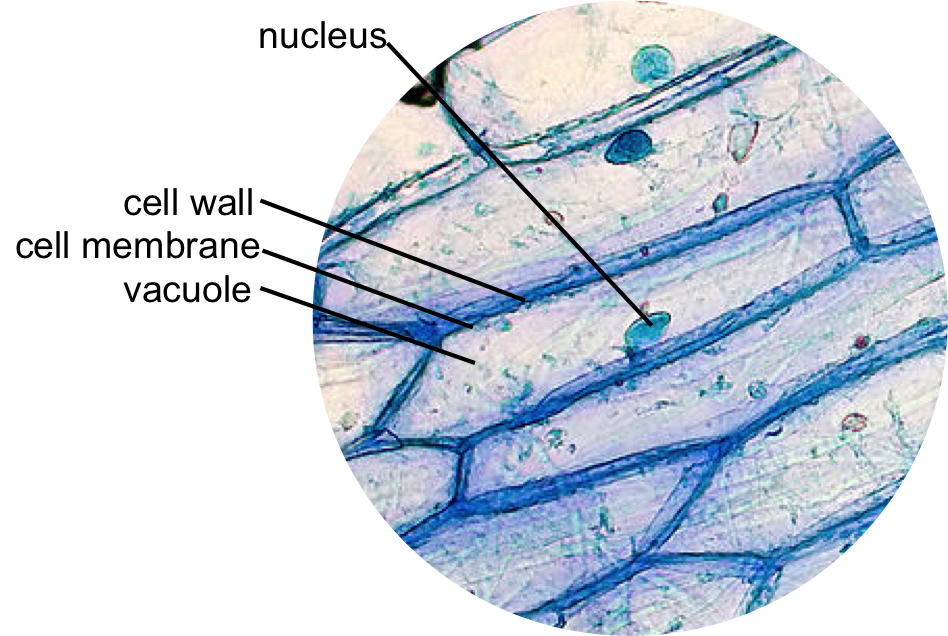
As we mentioned above iodine is the best stain to use when looking at onion cells. Observe an onion cell under the microscope.

Histolab4a Htm Histology Slides Science And Nature Animal Cell
Image will be uploaded soon.

. I like to buy supplies from Amazon and. Animal cell nucleus function plays the most important role for the cell. It was not until good light microscopes became available in the early part of the nineteenth century that all plant and animal tissues were discovered to be aggregates of individual cells.
There are various number of nuclei either they are single nucleus uni-nucleate two nuclei bi-nucleate or multi-nucleate. In this figure Cell wall provides additional protective layers outside the cell membrane. However the internal structure and organelles are more or less similar.
Most plant and animal cells are only visible under a light microscope with dimensions between 1 and 100 micrometres. The discovery of blood cells in the human body paved the way for advanced studies in cell biology. Aims of the experiment.
The cell from the Latin word cellula meaning small room is the basic structural and functional unit of lifeEvery cell consists of a cytoplasm enclosed within a membrane which contains many biomolecules such as proteins and nucleic acids. Once slides have been prepared they can be examined under a microscope. Life Sciences Under the Microscope Histology and Cell Biology.
That said there are other types of stains that can be used based on the type of cell that will be observed under the microscope and some of these can be used on onions as well. The nice thing about purchasing the appropriate supplies is that youll have them on hand the next time you want to use your microscope. Every animal cell does not have all types of organelles but commonly animal cells contain most of the organelles mentioned below.
Animal cells have different parts which contain many types of specialized organelles that help in carrying out various functions of the body. One observation was from very thin slices of bottle cork. Investigating cells with a light microscope.
Materials Needed for Observing Onion Cells. Animal cell under the microscope. Animal cells usually are transparent and.
Once slides have been prepared they can be examined under a microscope. A typical animal cell is 1020 μm in diameter which is about one-fifth the size of the smallest particle visible to the naked eye. The cell was first discovered by Robert Hooke in 1665 which can be found to be described in his book Micrographia.
Microscopes have contributed significantly in the fields of cell biology and histology where great discoveries have been made over the years. How to see the cell nucleus under a microscope. Aims of the experiment.
A typical animal cell is 1020 μm in diameter which is about one-fifth the size of the smallest particle visible to the naked eye. To use a light microscope to examine animal or plant cells. All the major organelles are mentioned below.
Since most cells are between 1 and 100 μm in diameter they can be observed by light microscopy as can some of. In order to examine cells in the tip of an onion root a thin slice of the root is placed onto a microscope slide and stained so the chromosomes will be visible. Microscopy plays a critical role in a majority of life sciences.
All eukaryotic cells have Nucleus few cells such as the mammalian RBCs may do not have. You will need to purchase some science materials for this lab exercise. Hooke discovered a multitude of tiny pores that he named cells.
A digital microscope or any simple light microscope. When looking under a microscope the cell wall is an easy feature to distinguish plant cells. Round is the most common shape of the nucleus but.
This discovery proposed as the cell doctrine by Schleiden and. In this book he gave 60 observations in detail of various objects under a coarse compound microscope. Investigating cells with a light microscope.
Under the microscope animal cells appear different based on the type of the cell. Here is how common. These regions of growth are good for studying the cell cycle because at any given time you can find cells that are undergoing mitosis.
Contemporary light microscopes are able to magnify objects up to about a thousand times. Animal cells do not have a cell wall. The light microscope remains a basic tool of cell biologists with technical improvements allowing the visualization of ever-increasing details of cell structure.
As a result most animal cells are round and flexible whereas most plant cells are rectangular and rigid. The cells youll be looking at in this activity were photographed with a light microsope. To use a light microscope to examine animal or plant.
There are two microscope lesson activities in this blog for you to see the nuclei in animal cells and plant cells. It is easier to see nuclei under a light microscope with staining such as methylene blue. Novel Coronavirus SARS-CoV-2 Under the Microscope The National Institute of Allergy and Infectious Diseases Rocky Mountain Laboratories NIAID-RML located in Hamilton Montana was able to capture images of the novel coronavirus SARS-CoV-2 previously known as 2019-nCoV on its scanning electron microscope and transmission electron microscopes.

Epidermal Onion Cells Under A Microscope Plant Cells Appear Polygonal From The Plant Cell Diagram Cell Diagram Plant Cell

List Of Cell Organelles Their Functions Plant And Animal Cells Animal Cell Cell Theory

Onion Epidermis Under Light Microscope Purple Colored Large Cells Project Microscopic Photography Microscope

Cell 8 Pictures Of Plant Cells Under A Microscope Plant Cell Structure Under Microscope Plant And Animal Cells Plant Cell Picture Plant Cell Structure

Animal Cell Organelles Sauna Design

Human Cheek Cell Wet Mount Under Phase Contrast Things Under A Microscope Cell Human

Scanning Electron Microscope Tumblr Scanning Electron Microscope Electron Microscope Electron Microscope Images

Plant Cell Under The Microscope 1 Microscopic Photography Plant Cell Microscopic Images

Year 11 Bio Key Points Cell Organelles And Their Function Cell Organelles Animal Cell Organelles

Microscopic Images Of Plant And Animal Cells Google Search Plant And Animal Cells Animal Cell Animal Cell Structure

Animal Cell Coloring Printing 5 Animal Cell Drawing In Cell Category Human Cell Diagram Animal Cell Drawing Cell Diagram

Cheek Cells Things Under A Microscope Cell Cheek

Plant And Animal Cells Revised Plant And Animal Cells Plant Cell Project Animal Cell Project

Animal Cell Cell Diagram Plant And Animal Cells

Structure Of Animal Cell And Plant Cell Under Microscope Diagrams Cell Diagram Plant And Animal Cells Plant Cell Diagram

What Is An Animal Cell Facts Pictures Info For Kids Students Animal Cell Drawing Animal Cell Animal Cell Anatomy

Cell Organelles Animal Cell Structure Organelles

Onion Epidermis With Large Cells Under Light Microscope Clear Epidermal Cells O Ad Microscope Light Epiderma Microscopic Cells Plant Cell Dna Project

Organic Cells Under Microscope Stock Photos Images Pictures 134 Images Plant Cell Cell Stock Photos